VIBT Imaging Center
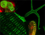
Although, the world around us mainly percepted through eyes, we forget the fact that our eyes are like small cameras, living optical devices whose lenses, drawing pictures on the retina, obey to the same physical laws which describe the behaviour of a polished piece of glass, the collecting lens. There is no comparable discovery of humanity like the collecting lens. The immense outer space and the infinite small can be viewed by lenses. Only by means of lenses, Galilei brought us the boundless universe and, Leeuwenhoek discovered living creatures not bigger than a few microns. The polished glasses really meant a revolution to the relationship of humans and their environment. With microscope lenses, one can have a closer look to nature and observe details which are usually out of sight and hidden from our busy daily life. Are you interested in exploring the „small world”? You can visit now the bioimaging core facility of our university. The long-desired microscope center has been established in the Muthgasse complex and harbours five high-end microscopes: two video microscopes, one of them dedicated to live cell imaging; the other one is designed for total internal reflection fluorescence microscopy, two confocal laser scanning microscopes and a laser microdissection microscope. Confocal microscopes are for better resolution in space, for example to study chromatin structure in nuclei. Video-microscopy, as its name suggests, serves as a camera to shoot movies from your samples. It provides excellent time resolution at the expense of some structural details. The live cell imaging platform is a hybrid system: one may use it like a video-microscope or, it can be transformed into a confocal one by attaching a laser scanning module to the stativ. Soon, an incubation chamber will complete the workstation to provide convenient growth temperature, CO2 and humidity for live samples. This system might become the favourite of neurologists and developmental biologists enabling them long lasting observations in natural conditions. With the arsenal of the imaging center students, professors, scientists are able to visualize, isolate and study fine structures of various objects and materials, not necessarily biological ones. It is possible to quantify and follow movements of microorganisms, organelles and even single molecules. Spatial and temporal changes of protein interactions, quantification of internal pH, redox potentials, concentration of ions, such as Calcium, and of molecules, such as glucose, will be possible to measure as long as their presence can be converted into fluorescent signals. Last but not least, one can feel the sensation of making surgery with a laser knife of the microdissecting microscope. This system allows you to separate and to collect specific regions of your sample, for example tumorous cells from tissues. By cutting a teeny-weeny part of a given specimen for further analysis, one may perform single cell DNA, RNA tests.
The imaging facility is open to all scientists and aims to initiate cooperations even beyond the BOKU. Welcome are cooperations to test innovative ideas and develop new microscopic methods.
- Invitation to the presentation on December 6th.
The center is headed by Dr Monika Debreczeny a physicist (MD and PhD in physics) with almost twenty years of experience in scientific research and running imaging laboratories in Hungary and in France. Do not hesitate to discuss your projects personally or by calling 47654 6951. You can find the BOKU-VIBT imaging center in Muthgasse 11, room A-UG02-O02 and the office of Dr. Monika Debreczeny is located in room 3-EG/G09. Within the first Quarter of the New Year, Monika wants to start the regular operation of the new facility with homepage, courses and booking system.